Damage to the fallopian tubes and the uterine lining
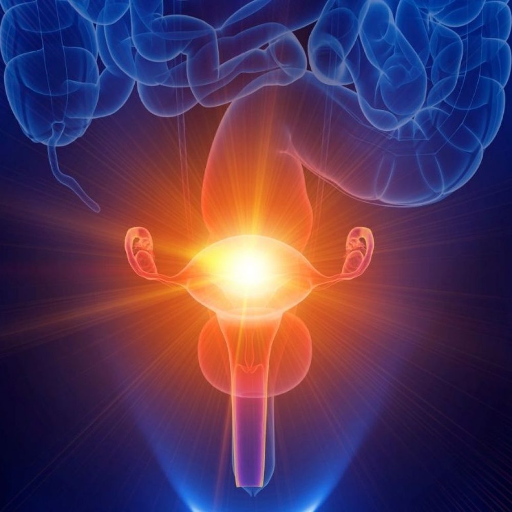
Damage can be caused by the inflammation caused by various sexually transmitted diseases. An inflammation can cause filmy adhesions to form around the fallopian tubes, ovaries or uterus that may interfere with the normal functioning of the female genitals. The formation of these adhesions can also be caused by surgeries in the pelvis minor region, including both intraperitoneal and intrauterine procedures. If the fallopian tube is damaged or closed, fertilisation of the egg is not possible due to a mechanical obstruction. Damage to the lining of the uterus, or endometrium, also prevents the embryo developed from a fertilised egg from attaching to the lining of the uterus and resulting in pregnancy.
Disorders of oocyte maturation and the ovarian release
Oocyte maturation in the ovaries is mainly mediated by two pituitary hormones: FSH (follicle-stimulating hormone) and LH (luteinising hormone). FSH induces the growth of the follicles needed by the egg, while LH helps the egg to mature and be released from the ovary (ovulation). Female infertility may occur due to an impaired regulation of FSH and LH. The most common hormonal disorder that causes female infertility is polycystic ovary syndrome (PCOS), in which the final maturation of the ovarian follicles is disrupted.
Endometriosis
Endometriosis is a condition where tissue similar to the lining of the womb starts to grow outsite of uterine cavity, such as the ovaries and fallopian tubes. Outbreaks of endometriosis cause damage to the ovaries and can result in a blockage of the fallopian tubes. The changes in the tissue fluids associated with endometriosis are detrimental to both the ova and very early embryos.
- Assess the levels of various hormones that can affect fertility (female and male hormones, thyroid hormones, prolactin, etc.) in the blood, and the relationships between those hormones. It is important to perform the blood test at the correct time – either on the 2nd or 3rd (maximum 5th) day of the cycle or on Day 21-22 during the daytime.
- Provide an analysis for various inflammatory agents. Tests are taken from the cervix.
- To provide a diagnosis of pre-tumorous conditions in the cervix, a so-called PAP analysis is required.
- A blood test is needed for the diagnosis of various viral diseases (HIV, hepatitis B and C, syphilis) and, if necessary, for autoimmune markers and the concentrations of vitamins.
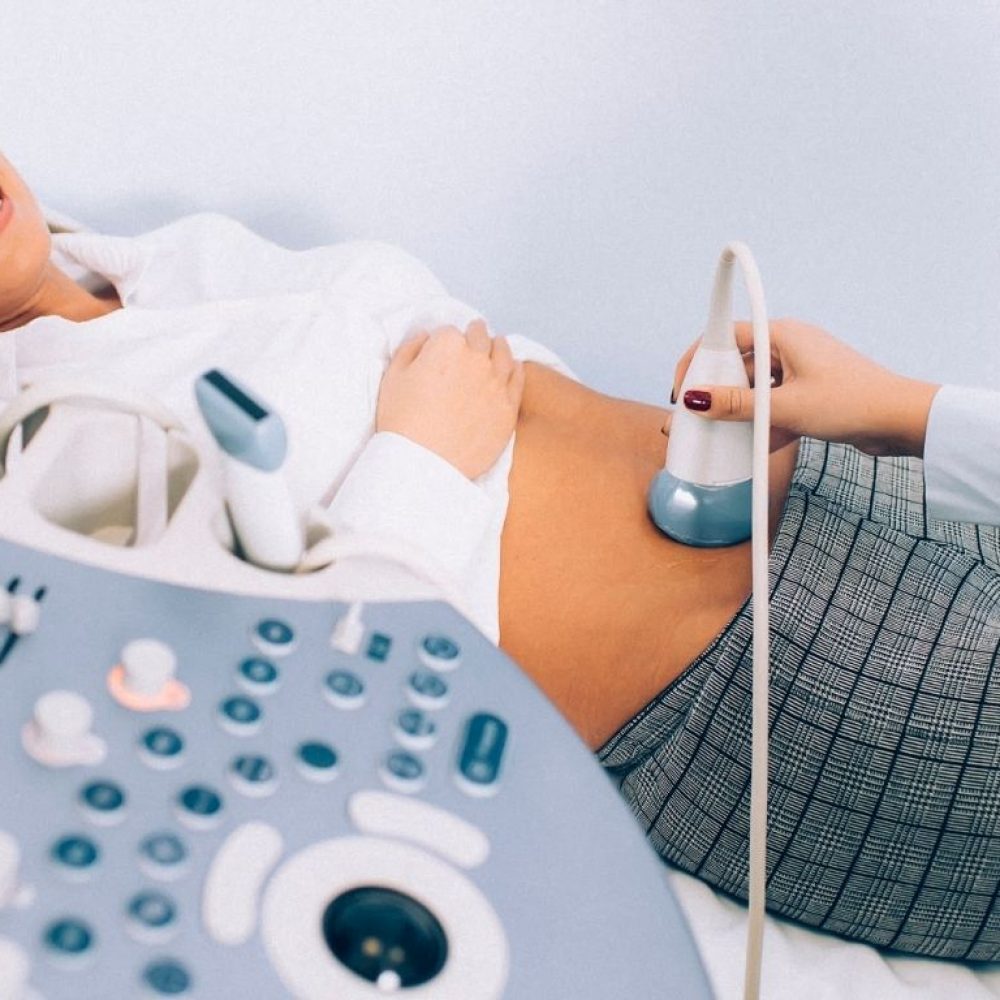
- An ultrasound is used to assess the uterine and ovarian anatomy and functions.
- A tubal permeability examination is performed either with a special contrast gel under ultrasound control (hysterosalpingo sonography) or, if necessary, as a laparoscopic surgery. Hysterosalpigo sonography is an outpatient procedure that does not require anaesthesia. It is recommended that the test be performed in the first half of the cycle, before ovulation takes place. During the procedure, a special gel is applied through the cervical canal to the uterus and fallopian tubes, with ultrasound monitoring in the areas where the gel is applied. However, the result of the study can be indirect, as the ultrasound does not reveal the presence of intrauterine adhesions.
- Laparoscopy is an operation that performed in the Department of Gynaecology under general anaesthesia. One must remain in the department for a period of 24 hours. Those with a history of pelvic inflammatory disease and suspected intrauterine adhesions may find the examination to be preferable, as intrauterine adhesions can be removed in addition to the tubal patency check. Endometriosis lesions can also be monitored and removed, if necessary. In addition, ovarian cysts or benign tumours of the uterus that may prevent pregnancy can be removed, if necessary.
- An examination of the uterus and uterine cavity is performed either with a special contrast gel under ultrasound control (hysterosonography) or, if necessary, through hysteroscopic surgery. Hysterosonography is an outpatient procedure that does not require anaesthesia. It is recommended that the test be performed in the first half of the cycle, before ovulation takes place. During the procedure, a special gel is inserted via cervix to the uterus and fallopian tubes, with ultrasound monitoring in the areas where the gel is applied. The examination generally reveals intrauterine polyps, the presence of myoma nodes, and their location in relation to the uterine cavity. The shape of the uterine cavity and its changes in the presence of congenital anomalies or intrauterine adhesions can also be determined. This study may be helpful in determining the need for surgery or in planning of the scope of the surgery. Hysteroscopic surgery can be performed either on an outpatient basis without anaesthesia or in the Gynaecology Department under general anaesthesia. Usually, the patient can go home on the same day. Hysteroscopy can be used to diagnose and, if necessary, treat intrauterine changes under specific visual controls.
- In the uterine mucosa i.e endometrial examination, a small sample of the uterine mucosa can be collected through a fine catheter and sent for a histological examination to determine the mucosal correspondence to the phase of the woman’s cycle or the presence of mucosal changes. The beREADY endometrial receptivity test The beREADY endometrial receptivity test is a modern personal medical test that takes the individual differences of each woman into account, and thus increases the effectiveness of the fertility treatment. According to studies, approximately 30% of patients require an individual approach and need to find a suitable time for an embryo transfer, because the uterine lining has not reached sufficient maturity by the scheduled embryo transfer date. The beREADY test evaluates the expression of the genes reflecting endometrial maturity by a high-precision DNA analysis. The result of this test gives the parties confidence that the embryo transfer will be performed at the most suitable time for the patient. More information is available at: beREADY.ee
- In addition, an endometrial receptivity evaluation test may be performed. For this test, specimens are taken from the endometrium, in a manner similar to that described above, from which the appropriate biomarkers are determined to assess the readiness of the endometrium for transplantation.
- In some cases, additional examinations by a geneticist and endocrinologist are indicated, such as magnetic resonance imaging for tumours, etc.